LIST OF MODELS THAT ARE MAPPED ON THE IMAGE – INSTRUCTIONS:
Instructions: Open each image by clicking on the name of the
link (in blue). After examining the
image, move your cursor over the structures or numbers (if present) to identify
them. The identity of the structure will show up on your computer screen if you hold the cursor over that structure
for a couple seconds.
I. SKELETAL SYSTEM &
ARTICULATIONS
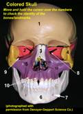 Colored Skull (anterior view)
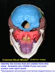 skull (colored, pull-apart; inferior view)
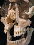 Facial bones from exploded skull (This model is not mapped, but should be used
for reference when studying individual bones of the face)
Facial bones from exploded skull (This model is not mapped, but should be used
for reference when studying individual bones of the face)
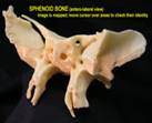
 Ethmoid bone (antero-lateral view)
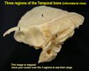 Temporal bone
(infero-lateral view)
Temporal bone
(infero-lateral view)
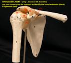 shoulder joint & ligaments (anterolateral
view)
shoulder joint & ligaments (anterolateral
view)
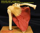 Shoulder joint with muscles attached
(anterior view)
Shoulder joint with muscles attached
(anterior view)
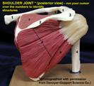 Shoulder
joint with muscles attached (posterior view)
Shoulder
joint with muscles attached (posterior view)
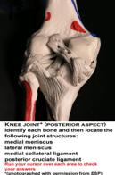
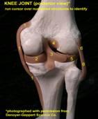 Knee joint model (posterior view)
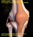
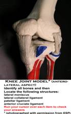 Knee joint (anterolateral view)
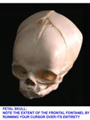 fetal skull – frontal fontanel
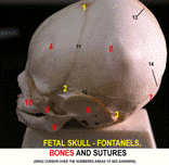 Fetal Skull (lateral view) (fontanels are labeled in yellow, bones in
red and sutures in black- you must place cursor over the suture itself (not
necessarily the number)
Fetal Skull (lateral view) (fontanels are labeled in yellow, bones in
red and sutures in black- you must place cursor over the suture itself (not
necessarily the number)
to see its identity